Tuesday, August 18, 2020

Proposed experiment with the genus Alexandrium catanella

Paralytic shellfish toxins (PSTs) constitute a suite of harmful neurotoxins commonly produced in marine ecosystems by several species of dinoflagellates within the genus Alexandrium.  Dinoflagellates  are eukaryotes and are usually considered algae. Most dinoflagellates are marine plankton There are up to 30 species of dinoflagellates in the genus Alexandrium. Species are defined by morphological features found in their thecal plate. Thecal plates are composed of cellulose or polysaccharide microfibrils. The size, shape and arrangement of thecal plates are used to distinguish dinoflagellate species. Light microscopy is used to find and determine the presence of Alexandrium. However, scanning electron microscopy is used to characterize the thecal plates and distinguish one species from another.

Documented cases of PSP in Alaska date back centuries to Captain George Vancouver’s survey of the Pacific coast in the early 1790s (Quayle 1969; Horner et al.1997).

Vandersea et al. (2017) not only developed new assays for the Alexandrium species most likely to be present in Alaska they also did a systematic evaluation of all published (~150) Alexandrium species- specific assays. They collated published Alexandrium PCR, qPCR, and in situ hybridization assay  primers and probes that targeted the small-subunit (SSU), internal transcribed spacer (ITS/5.8S), or D1–D3 large-subunit (LSU) (SSU/ITS/LSU) ribosomal DNA genes.

Garneau et al. (2011)  used molecular beacon-based qPCR method to detect and quantify A. catenella in pure cultures and in mixed natural plankton assemblages.

 Proposed experiments:

Culture Alexandrium species. Cell lysates created from A. catenlla culture (ACRB01) of known abundance will be used to correlate cell numbers to qPCR threshold cycles (Ct) using a standardized protocol. There are variances in copy number within dinoflagellates. So, a calibration curve of 5 point 1:10 serial dilution of a known abundance of actively growing A. catellena cells. Cell numbers will be determined by light microscopy. Primers UScatF and UScatR and UScatMB with 6-fluorescein amidite (6-FAM) at the 5' end (synthesized by Eurofins MWG Operon) will be used for qPCR.

In an effort to predict toxic PST events, we will collect A. catenlla samples along with salinity, temperature, nutrients, chlorophyll, and PST biotoxin levels. Data from three marine stations in Bellingham Bay, WA will be documented in order to determine which environmental conditions promote the onset and flourishing of PST events or unusual bloom events.

References:

Quayle, Paralytic Shellfish Poisoning in British Columbia, Fisheries Research Board of Canada; First Edition edition (1969)

Galluzzi, L., et al. (2004) Development of a real-time PCR assay for rapid detection and quantification of Alexandrium minutum (a dinoflagellate). Appl. Environ. Microbiol. 70:1199–1206.

Moorthi, et al. (2006) Use of quantitative real-time PCR to investigate the dynamics of the red tide
dinoflagellate Lingulodinium polyedrum. Microb. Ecol. 52:136–150.


Garneau et al. (2011) Examination of the Seasonal Dynamics of the Toxic Dinoflagellate Alexandrium catenella at Redondo Beach, California, by Quantitative PCR

Vandersea et al. (2017) qPCR assays for Alexandrium fundyense and A. ostenfeldii (Dinophyceae) identified from Alaskan waters and a review of species-specific Alexandrium molecular assays

Monday, August 17, 2020

Taqman PCR for quantification of microcystin production (after Kurmayer and Kutzenberger, 2003)

Microcystis cause freshwater cyanobacteria blooms that effect drinking water, water-based recreation, and ecology. 

https://www.usgs.gov/media/images/microcystis-aeruginosa-bloom-0

Light microscopy image from Wikipedia

Microcystis wesenbergii colony under epifluorescence microscopy with SYTOX Green DNA staining. Image from Wikipedia.

Microcystis produce microcystin.


Microcystin is a nonribosomally synthesized cyclic hepatotoxin (liver toxin) with potent inhibitory activity against mammalian protein phosphatases.


Recently, the TaqMan PCR, or the Taq nuclease assay (TNA), was introduced to quantify specific genotypes of picocyanobacteria (Becker et al., 2000) or microcystin-producing cyanobacteria in the field (Foulds et al., 2002). 

TNA utilizes a sequence specific dually labeled fluorescent probe (TaqMan probe) and primers to quantify the level of DNA template initially present in a sample.
 

The rate of exponential accumulation of the amplicon is monitored by the hydrolysis of the TaqMan probe, in which it generates a fluorescent signal during the amplification process.


The threshold cycle (Ct) is the PCR cycle number at which the fluorescence passes a set threshold level and can be used to determine the starting DNA amount in the sample based on a standard curve (based on samples with a known concentration).

For Kurmayer and Kutzenberger (2003), cell numbers inferred from TNA standard curve correlated significantly with the microscopically determined (particle counting) cell numbers on a logarithmic scale. 

They conclude that the Microcystis cell numbers could be used to infer the mean proportion of mcy genotypes in Lake Wannsee (Berlin, Germany). I could use these methods to study mcy genotypes in Bellingham Bay. Kurmayer and Kutzenberger (2003) do not mention issues with multiple copy numbers or cross-reactivity. 

 Proposed experiments to study Microscystis:

Culture several unicellular strains of Microcystis sp. to test primer and TaqMan probe sensitivity and specificity. TaqMan PCR, or the Taq nuclease assay (TNA) will be used to quantify the mcyB region. The TaqMan probes are from Kurmayer and Kutzenberger (2003; 5' - CACCAAAGAAACACCCGAATCTGAGAGG-3) The probe will have a fluorescent reporter dye (6-carboxyfluorescein) covalently attached to the 5' end (5' -FAM) and a 3' -TAMRA fluorescent quencher
dye (6-carboxytetramethylrhodamine). A standard curve based on predetermined cell concentrations will be established by relating the known DNA concentrations (in cell equivalents) to the Ct of the diluted samples.

References:

Becker et al., (2000) PCR Bias in Ecological Analysis: a Case Study for Quantitative Taq Nuclease Assays in Analyses of Microbial Communities

Foulds et al., (2002) Quantification of microcystin-producing cyanobacteria and E. coli in water by 5    -nuclease PCR

Kurmayer and Kutzenberger (2003) Application of Real-Time PCR for Quantification of Microcystin Genotypes in a Population of the Toxic Cyanobacterium Microcystis sp.

Science and Family pt. 2

In a few days my mom, dad, my son, and I will be moving to Everson, WA. I started a new postdoc at the Salish Sea Research Center. Our belongings are now on a truck and on their way to Washington State.

One year ago my husband moved to State College to start medical residency. Previous to this he was separated from my son and me. This is because three years ago I started my first postdoc at Penn State University. Now, we are separating again. Hopefully, for the last time in our lifetime.

In the meantime, my mom and dad continue to travel with me on this academic journey. They are raising my son while I pursue science as a career. My mom is a teacher and she is homeschooling my son this year. He will hopefully start Kindergarten next fall. We are moving into a cabin on 5 acres. We are all very excited to start this next phase. We are also very sad to leave my husband and our house in State College, PA.

My son, my mom, my dad in Coburn, PA ( January 2019).

Science and Family pt. 1

My husband and I attended the 2011 Geological Society of America meeting in Minneapolis, MN. This is us posing in front my poster.
 
My husband, my son, and I posing in front of beamline 12.3.2 at the Advanced Light Source in 2016.

Anabaena planktonica

Anabaena planktonica is a harmful, bloom-forming freshwater cyanobacterium.

 Anabaena planktonica


Pseudo-nitzschiamarine rbcS qRT-PCR assay

 Pseudo-nitzschiamarine, a genus of diatom, includes species that produce domoic acid, a neurotoxin
responsible for illness and mortality in both humans and marine wildlife. 

Pseudo-nitzschia has been identified as one of the main genera to increase in cell numbers during iron fertilization experiments in high nitrate low chl (HNLC) regions of the Southern Ocean, the subarctic Pacific, and the east equatorial Pacific (de Baar et al. 2005). 

https://en.wikipedia.org/wiki/Pseudo-nitzschia#/media/File:Pseudonitzschia2.jpg
 

Although no additional human cases have been confirmed, bioaccumulation of domoic acid has resulted in illness and mortalities of many birds and marine mammals, especially along the Pacific coast of North America (Gulland, 2006; Scholin et al., 2000; Sierra-Beltra´n et al., 1997; Work et al., 1993). 

Domoic acid

https://caseagrant.ucsd.edu/project/frequently-asked-questions-domoic-acid-in-california-crabs
 

The threat posed by human consumption of contaminated shellfish has led to periodic closures of commercial and recreational harvesting areas, resulting in significant economic
losses around the world (Gallacher et al., 2001; Trainer, 2002; Tweddle et al., 2010).  

The genus Pseudo-nitzschia is composed of at least 30 species and is common to diatom assemblages in all ocean basins, both in coastal and offshore waters.  Morphological species complexes and/or cryptic species are increasingly described for the genus (Hasle 1995, Manhart et al. 1995, Villac and Fryxell 1998, Lundholm et al. 2002, 2003, 2006, Orsini et al. 2004, Hasle and Lundholm 2005).

Molecular methods provide early detection of blooms Pseudo-nitzschiamarine and predict toxin accumulation.

Delaney et al. (2011) developed a quantitative reverse transcription PCR (qRT-PCR) assay for the detection of ribulose-1,5-biphosphate carboxylase/oxygenase small subunit (rbcS) gene. 

The rbcS qRT-PCR assay is useful for the detection and enumeration of low concentrations of P. multiseries in the environment.

Nucleic acids

The nucleic acids, the vital constituents of living beings, are long-chain polymers composed of nucleotides. Nucleic acids were named based partly on their chemical properties and partly on the observation that they represent a major constituent of the cell nucleus. The fact that they form the chemical basis for the transmission of genetic traits was not realized until 1941. Among other important roles, nucleotides can serve as sources of energy in the form of ATP, physiological signaling mediators, secondary messengers, and allosteric enzyme effectors.

Effects of eutrophication of ecosystems

Anthropogenic activities are increasing. Activities include urban, agricultural, and industrial land development. The activity has resulted in the eutrophication of numerous fresh-water ecosystems worldwide. The excess of nutrients, such as phosphorus and nitrogen increase the growth rate of phytoplankton resulting in the formation of dense populations (i.e., algae blooms). 

Algae blooms, cyanobacterial blooms, have been shown to pose environmental and social
problems (Vargas-Montero and Freer 2004; Haande et al. 2007; Stone and Bress 2007; Zhang et al. 2007). 

Blooms reduce water quality and the recreational value of aquatic ecosystems (Rahman at al. 2005; Smith and Lester 2006).

 Cyanobacterial species produce potent toxins, which pose serious health risks to both human and animals (Codd et al. 2005).

Tuesday, August 11, 2020

qPCR


Quantitative real-time PCR technologies (what is Taqman qPCR):


https://www.ncbi.nlm.nih.gov/probe/docs/techqpcr/
Quantitative real-time PCR (qrt-PCR) is a good option to generate ‘real-time’ data regarding the presence of harmful algal species. The major goal of a qrt-PCR project is to design and test a qrt-PCR assay that will accurately identify and estimate marine algal species. 

Quantification is performed during the exponential phase of the PCR, where amplification efficiency is maximum. In real-time PCR, amplicon formation is monitored after each cycle by measuring a fluorescence signal.

An amplicon is a piece of DNA or RNA that is the source and/or product of natural or artificial amplification or replication events. It can be formed using various methods including polymerase chain reactions (PCR) or natural gene duplication.

In qrt-PCR, the increase in fluorescence observed during the reaction will be proportional to the starting quantity of the target molecule. Fluorescence can be generated by using fluorescent probes such as TaqMan®. Since there is a correlation between the cycle number at which the amplicon is initially detected (threshold cycle, Ct) and the starting amount of target molecules, it is possible to calculate the amount of target sequence in an unknown sample by using a standard reference curve generated using DNA extracted from a known number of cultured cells. 

Monday, August 10, 2020

Postdoctoral Molecular Researcher position at Northwest Indian College


As a Postdoc and Molecular Researcher at Northwest Indian College on the Lummi main campus at the Salish Sea Research Center (SSRC), I will monitor harmful agal bloom species (Alexandrium catenella, Azadinium poporum, and Pseudo-nitzschia multiseries) in the Bellingham and Lummi Bays (Washington State) using molecular techniques. 

Protocols for monitoring harmful algal bloom species will used in inform the local Lummi Nation. This project will provide food and data sovereignty for the Lummi Nation and Lummi Natural Resources deparment about annual harmful algal blooms. 

Gooseberry Point along the Bellingham Bay


Friday, August 7, 2020

Career update for years 2008-2020

Background:

LSAMP-Bridge to the Doctorate cohort VI.

As I have not updated this blog since I was a Master's student at the University of New Mexico (Image above taken in 2008) working with Professor Laura Crossey on the biogeochemistry of the Tierra Amarilla Anticline in northern New Mexico, I thought it was a good idea to catch up on my background. As a biogeochemist, I have used and become an expert in a variety of different imaging and spectroscopy tools. 


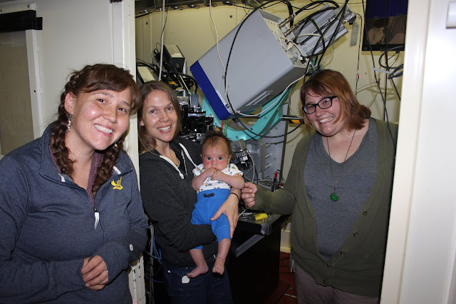
(from left to right) Brandi Cron, Professor Brandy Toner, Robert Atticus Kamermans and Aubrey Dunshee taking a photo break with Robert Atticus Kameramans at BL 12.3.2 at the Advanced Light Source.

As a Ph.D. candidate at the University of Minnesota (image above taken in 2017), I gained expertise characterizing abiotic and biotic mineral precipitates in deep-sea hydrothermal vents, using Scanning Transmission X-ray Microscopy, X-ray Absorption Near Edge Structure (XANES) and X-ray diffraction.

Pennsylvania State University geosciences professor Julie Cosmidis and postdoctoral fellow Brandi Kamermans prepare to change out samples on the SM beamline. Photo is from the Canadian Light Source.

Most recently, I have been using Scanning Transmission X-ray Microscopy, Raman, and Scanning Electron Microscopy at the Pennsylvania State University Material Characterization Laboratory to characterize both elemental sulfur and organics produced by Sulfuricurvum kujiense, in an effort to distinguish chemical versus microbial induced mineral production.